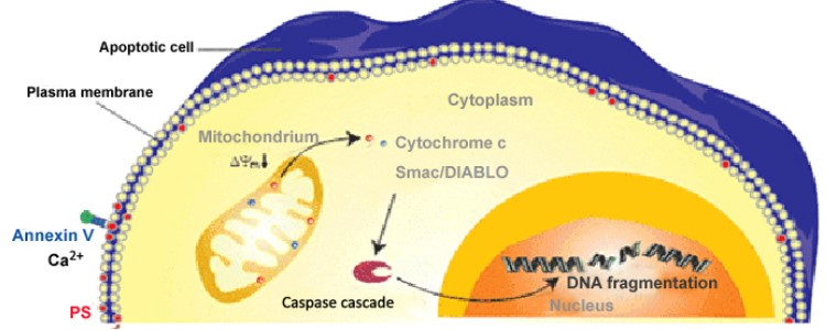
細胞凋亡(Apoptosis)是指細胞為維持細胞內環境穩定,由基因控制的細胞自主性有序的死亡。 在細胞凋亡發生期間,細胞膜、線粒體、細胞質及細胞核都發生了不同程度的變化,這些變化都可以作為評估細胞凋亡的依據。在凋亡發生的最初時期,細胞膜上的磷脂醯絲氨酸(phosphatidylserine,PS) 由細胞膜內側外翻至細胞膜外表面。接著線粒體隨著其膜電位的改變開始去功能化,釋放出一些線粒體蛋白,如細胞色素C(Cytochrome c)和Smac蛋白,由此導致了下游的caspase啟動和DNA的片段化發生。
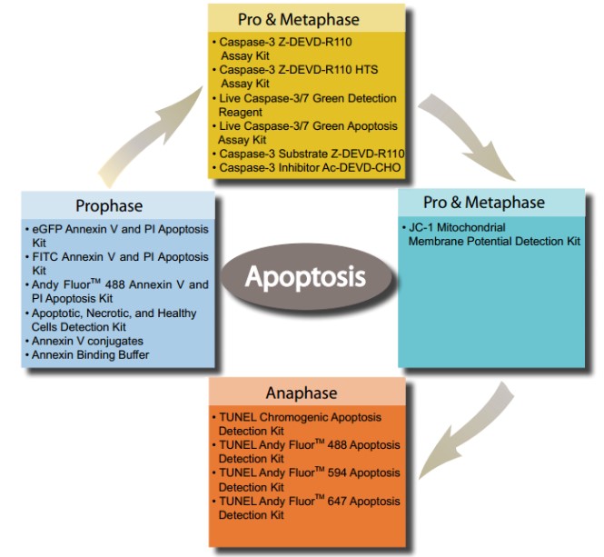
ABP Biosciences提供一系列檢測細胞凋亡的套組,測量細胞凋亡過程中不同階段的各種指標。
- Annexin V binding assays
- Caspase activity assays
- Mitochondrial membrane potential assays
- TUNEL assays
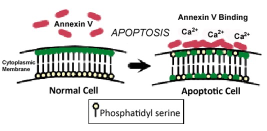
Annexin V binding assays
Annexin V是35–36 kilodalton, Ca2+ -dependent磷脂結合蛋白,其對磷脂醯絲氨酸(phosphatidylserine,PS)具有高親和力。 在正常存活的細胞中,PS位於細胞質內的細胞膜表面。在凋亡早期,細胞PS從質膜的內部轉移到外部,使PS暴露細胞外部環境,可通過annexin V conjugate檢測暴露於細胞外的PS。更多詳情>>
- eGFP Annexin V and PI Apoptosis Kit
- FITC Annexin V and PI Apoptosis Kit
- Andy Fluor™ 488 Annexin V and PI Apoptosis Kit
- Apoptotic, Necrotic, and Healthy Cells Detection Kit
- Annexin V Conjugates
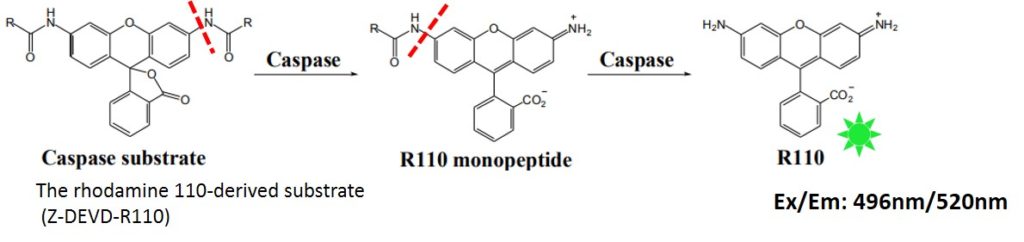
Caspase activity assays
細胞凋亡早期階段的一個顯著特徵是caspase enzymes的活化,其參與蛋白質的切割和隨後的細胞分解。其中caspase-3的啟動在凋亡信號傳導的許多途徑中發揮著關鍵的作用。活化的Caspase-3可以剪切特定的氨基酸序列。針對這種特性,GeneCopoeia提供了具有高度特異性Caspase-3/7螢光染劑用來檢測Caspase-3/7 活性。 更多詳情>>
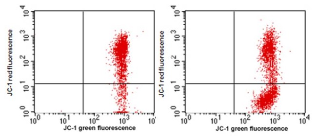
Mitochondrial membrane potential assays
JC-1在正常的cell中會聚集在mitochondria內,形成J-aggregates發出紅色螢光。而在Apoptotic cell中mitochondria膜電位改變,JC-1無法堆積在mitochondria中而會以單體形式留在cytoplasm中發出綠色螢光。更多詳情>>
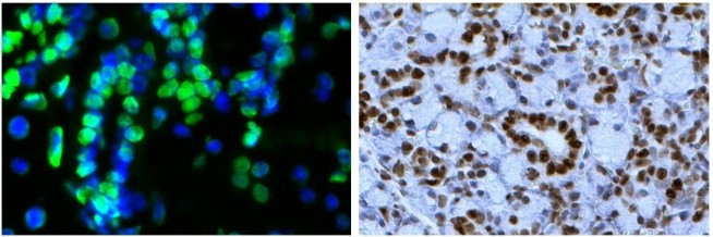
TUNEL assays
細胞凋亡的後期階段的特徵是細胞核產生形態學的變化,包括DNA片段化、染色質濃縮、核膜退化、核變色和DNA鏈斷裂。 在凋亡期間發生的DNA片段化產生DNA鏈斷裂,其可以使用TUNEL (terminal deoxynucleotidyl transferase dUTP nick end-labeling) 測定法進行分析。更多詳情>>
- TUNEL Chromogenic Apoptosis Detection Kit
- TUNEL Andy Fluor™ 488 Apoptosis Detection Kit
- TUNEL Andy Fluor™ 594 Apoptosis Detection Kit
- TUNEL Andy Fluor™ 647 Apoptosis Detection Kit